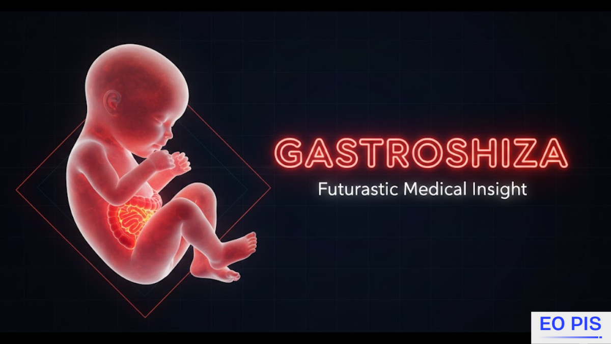
Gastroshiza is a rare congenital condition that affects newborns and is defined by an abdominal wall defect that forms early in fetal development. Because the abdominal wall does not completely seal, parts of the intestine and sometimes other organs become internal organs exposed, leading to abdominal organs outside the body. This visible protrusion of abdominal organs through a hole in the abdominal wall marks the condition as a serious congenital disorder within the group of gastrointestinal abnormalities.
In many clinical notes,, the same condition is also written as gastrophiza, but the underlying defect is the same. Management requires coordinated care from neonatologists, pediatric surgeons, and genetic counselors, especially in hospitals with strong neonatal intensive care facilities.
What is gastroshiza?
In gastroshiza, the defect is typically an abdominal wall defect near the umbilical cord where the bowel emerges directly into the amniotic fluid. At birth, the clinician sees abdominal organs outside the body, often with intestines protruding to the right of the umbilical cord. Because the organs are internal organs exposed without a protective sac, they are vulnerable to irritation, infection, and later long-term gastrointestinal problems.
Some babies have larger abdominal wall defects with the involvement of the stomach or liver, or additional anomalies such as congenital heart defects and chromosomal abnormalities. These cases demand more complex treatment planning and stricter monitoring.
Despite the severity, outcomes have improved because of prenatal imaging methods, early diagnosis, and structured long-term management plans guided by continuous pediatric support.
Causes and Risk Factors of gastroshiza
The exact causes of gastroshiza are not fully defined, but evidence points to a mix of genetic predisposition and environmental factors in pregnancy. A subtle inherited component combined with specific external triggers seems to disrupt normal fetal development of the abdominal wall.
On the genetic side, minor genetic mutations affecting blood flow or tissue formation may contribute to the defect. Families with a strong family history of congenital disabilities often benefit from genetic counseling before or during pregnancy to understand risks and available tests,, such as amniocentesis.
Environmental and maternal factors add further risk:
- maMaternalxposure to tobacco
- maternal alcohol use
- certain medications in early gestation
- nutritional deficiencies and lack of essential nutrients in pregnancy, especially folic acid, before and during pregnancy
Broader social factors also matter. Socio-economic status can limit diet quality and access to prenatal care and regular check-ups, leading to limited healthcare access and weaker nutrition education. These conditions make it harder to maintain a well-balanced diet in pregnancy and to identify problems early.
Genetic predisposition and environmental factors in pregnancy
Most cases are sporadic, but a combination of genetic predisposition and environment shapes risk. A fetus with a mild inherited component may be more vulnerable to harmful environmental factors in pregnancy, like maternal exposure to tobacco, maternal alcohol use, inappropriate medications in early gestation, and severe nutritional deficiencies.
Improving maternal health and behavior—through avoiding smoking and alcohol, avoiding harmful drugs and chemicals, using folic acid before and during pregnancy, and maintaining a healthy pre-pregnancy weight—helps reduce overall risk and also decreases obesity-related pregnancy complications.
Symptoms and Early Detection
Clinically, gastroshiza is obvious at birth because of the protrusion of abdominal organs and the unmistakable abdominal organs outside the body. But modern practice relies heavily on early detection in pregnancy using prenatal imaging methods.
Routine screening with routine prenatal ultrasounds often includes an 18–20 week ultrasound. During this exam, ultrasound scans or simply an ultrasound scan can show bowel floating outside the abdomen, indicating an abdominal wall defect. This early view allows better treatment planning and delivery at a center with neonatal intensive care.
After birth, functional symptoms are common:
- Feeding difficulties after birth
- swallowing and digestion problems
- signs of gastrointestinal blockage
- increased risk of post-delivery infection
- possible respiratory issues and impaired lung development in severe cases
These findings drive urgent postnatal evaluations and rapid preparation for surgery shortly after birth.
Diagnosis of gastroshiza
Diagnosis usually unfolds in two phases: prenatal and postnatal.
During pregnancy, prenatal imaging methods such as ultrasound scans are key. A standard 18–20 week ultrasound often reveals the defect. If bowel loops are seen outside the abdomen at an abdominal wall defect near the umbilical cord, clinicians suspect gastroshiza.
When additional anomalies are suspected, amniocentesis helps look for chromosomal abnormalities, and focused cardiac imaging checks for congenital heart defects.
After delivery, diagnosis is confirmed by a direct physical examination after birth. The team assesses:
- Size of the defect and any larger abdominal wall defects
- degree of involvement of the stomach or liver
- color, swelling, and viability of bowel
- The overall extent of organ involvement
Additional imaging studies, such as X-rays and CT sca,,ns refine understanding of bowel position, possible gastrointestinal blockage, and guide detailed treatment planning.
Diagnostic Procedures
Key diagnostic tools include:
- ultrasound and ultrasound scans as first-line prenatal imaging methods
- amniocentesis in selected cases with suspected chromosomal abnormalities
- postnatal X-rays to look for bowel obstruction and tube placement
- CT scans are used in atypical or complex anatomy to define the extent of organ involvement.
These steps help the team of neonatologists, pediatric surgeons, and genetic counselors design a safe path to abdominal cavity repair.
Treatment Options for gastroshiza
Treatment centers on protecting the internal organs exposed, stabilizing the baby, and performing abdominal cavity repair. In most cases, surgery shortly after birth is required.
Immediately after delivery, the baby is moved to a neonatal intensive care unit or neonatal intensive care unit (NICU), where neonatal intensive care focuses on:
- Covering abdominal organs outside the body to reduce the risk of infection
- Giving intravenous fluids for stabilization
- careful monitoring of vital signs
- Beginning nutrition management with IV nutrition until the bowel tolerates feeds
Surgical repair involves repositioning protruding organs into the abdomen and closing the defect. Depending on swelling and space, the team may choose direct closure or staged surgeries.
Immediate Postnatal Care
Early hours in the neonatal intensive care unit are critical. Staff apply sterile coverings, start intravenous fluids, manage nutrition management, and use continuous monitoring of vital signs to watch for respiratory issues, risk of infection, and circulatory instability.
Only after this stabilization does the team move to the operating room for early surgical intervention.
Surgical Intervention
If feasible, surgeons perform one-step abdominal cavity repair by repositioning protruding organs and closing the abdominal wall defect. When tension is too great, they use staged surgeries with a silo and gradual closure of the abdominal wall over several days. Throughout, they try to apply scar minimization techniques without compromising safety.
Postoperatively, babies stay in the neonatal intensive care unit (NICU) under neonatal intensive care, progressing through:
- Continued intravenous fluids and nutrition management
- slow transition to oral feeding
- Possible specialized feeding techniques if there are swallowing and digestion problems
- watching for bowel obstruction, post-delivery infection, and the need for repeat surgery
Long-term, many children require dietary modifications, physical therapy, and structured long-term management plans.
Complications Associated with gastroshiza
Even with expert care, gastroshiza can lead to several complications:
- Risk of infection and post-delivery infection due to the internal organs exposed
- Malnutrition in infants occurs when feeding difficulties occur after birth or prolonged IV feeding limits intake. Bowel obstruction and other long-term gastrointestinal problems that may need repeat surgery
- respiratory issues and impaired lung development in infants with tight closures or large defects
- developmental delays from extended hospital stays and critical illness
- Ongoing emotional and psychological challenges and strong parental anxiety around surgeries and follow-up
These outcomes justify close regular follow-up care and early intervention services.
Prognosis and Long-Term Outcomes
With current practice, prognosis and survival rates for gastroshiza are generally good. Many babies survive and ultimately eat, grow, and develop well, especially when early surgical intervention is combined with disciplined long-term management.
Important factors include:
- Degree and extent of organ involvement
- presence of congenital heart defects or chromosomal abnormalities
- severity of early complications such as bowel obstruction or severe infection
- access to continuous pediatric support and structured regular follow-up care
Most children reach positive long-term outcomes with appropriate dietary modifications, physical therapy, and educational support if developmental delays occur.
Preventive Measures and Maternal Care
No strategy can prevent every case, but several measures reduce risk:
- Consistent prenatal care and regular check-ups with routine prenatal ultrasounds and other prenatal imaging methods
- a well-balanced diet in pregnancy rich in essential nutrients in pregnancy, including folic acid before and during pregnancy, and folate-rich foods (leafy greens, legumes, fortified cereals)
- avoiding smoking and alcohol, and avoiding harmful drugs and chemicals to minimize harmful environmental factors in pregnancy
- Maintaining a healthy pre-pregnancy weight and lowering obesity-related pregnancy complications through moderate exercise in pregnancy
Improving socio-economic status at the community level, reducing limited healthcare access, and strengthening nutrition education programs all support better outcomes for mothers and babies, including those at risk of gastrointestinal abnormalities like gastroshiza.
Living with gastroshiza
Life after discharge involves ongoing long-term management rather than a simple cure. Families coordinate with continuous pediatric support to track growth, behavior, and school progress. Children may live with residual long-term gastrointestinal problems or mild respiratory issues and may need intermittent dietary modifications, specialized feeding techniques, and physical therapy.
For parents, the constant appointments, monitoring for bowel obstruction, and fear of repeat surgery create significant emotional and psychological challenges and persistent parental anxiety. Many families also juggle coping with chronic medical conditions alongside work and other children.
As children improve, fostering independence in children becomes essential: encouraging them to manage minor symptoms, participate in school and activities, and understand their history without feeling defined by it.
Supporting Families and Resources
Families benefit from structured social and emotional support:
- Support groups for families offer shared experience and practical strategies for coping with chronic medical conditions linked to gastroesophageal.
- Hospital programs can strengthen nutrition education and guide parents through specialized feeding techniques and home care.
- Social services can help reduce the impact of limited healthcare access and financial strain tied to repeated hospital stays and follow-up.
With informed care, consistent regular follow-up care, and strong support systems, children with gastroshiza and gastrophiza variants can achieve stable long-term outcomes, and families can gradually regain control over daily life.
Conclusion
Gastroshiza is a rare congenital condition and serious congenital disorder marked by an abdominal wall defect through a hole in the abdominal wall, leaving internal organs exposed and sometimes significant abdominal organs outside the body. Despite its severity, modern prenatal imaging methods, reliable routine prenatal ultrasounds, and proactive early detection in pregnancy allow better treatment planning, delivery at centers with strong neonatal intensive care, and timely surgery shortly after birth. Outcomes depend on the extent of organ involvement, presence of associated congenital heart defects or chromosomal abnormalities, and the quality of long-term management—including dietary modifications, vigilant surveillance for bowel obstruction, and readiness for possible repeat surgery.
FAQs
What exactly is gastroschisis?
Gastroschisis is a birth defect where the baby’s intestines protrude through a hole in the abdominal wall.
Can a baby survive gastroschisis?
Yes, most babies survive with early surgical intervention and neonatal care.
What is the treatment for gastroschisis?
Treatment involves surgery to reposition the exposed organs and close the abdominal wall defect.
Which drug causes gastroschisis?
No single drug is proven to cause gastroschisis, but certain medications increase the risk.
Is gastroschisis a high-risk pregnancy?
Yes, gastroschisis is considered a high-risk pregnancy due to potential complications.
What medications increase the risk of miscarriage?
Medications like NSAIDs, methotrexate, and some hormonal treatments may increase the risk of miscarriage.